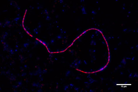
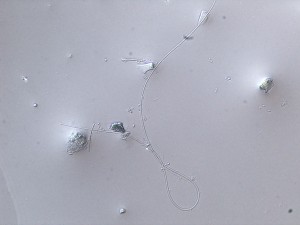
Fluorescence In Situ Hybridisation (FISH) is a technique to detect and localize the presence of specific micro-organisms within an environmental sample. FISH uses specifically designed fluoresent probes that bind to the ribosomal RNA of specific bacteria. Fluorescence microscopy is used to find out which micro-organisms are labelled. In 2013, as part of the ERC project, a new laboratory for FISH was installed at NIOZ Yerseke. To kick start this new facility, a 4 day training course on FISH was organized, with lectures by Dr. N.M. Lee (Department Microbiology, TU Muchen), a leading international expert on FISH. Furthermore, Silvia Hidalgo Martinez joined the team as Technical Support Assistant specifically dedicated to FISH analysis. The FISH technique is crucial to confirm the indentity and quantify the abundance of Electrogenic filaments in our sediments. Silvia spent a month at the Department of Bioscience (Center for Geomicrobiology, University of Aarhus in Denmark), a research group with whom we have a strong collaboration. In Aarhus, Silvia received a dedicated training by Dr. Regina Schauer, the international specialist on FISH analysis of Electrogenic filaments. The image enclosed shows an electrogenic filamentous bacterium stained with both a general DNA stain and a specific FISH probe (red stain, DSB706 probe for Desulfobulbaceae).