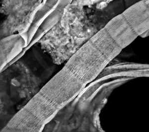
Figure 1. Scanning electron microscopy image of the external surface of a cable bacterium filament. The surface shows a set of parallel ridges that run along the whole length of the filament. These ridges harbour the conductive structures.
A record breaking conductivity
Scanning electron microscopy reveals that the outer surface of the cable bacteria has an unusual topography, with parallel ridges that run along the axis of the cable bacteria (Figure 1). Novel observations using state-of-the-art electron microscopy and atomic force microscopy provide a detailed picture of these ridge structures (Cornelissen et al., 2018). Cryo-transmission electron tomography shows that fiber structures are embedded in the cell envelope underneath the ridges. Combining different types of microscopy data we have made a full 3D model of how the fiber network is embedded in the cell envelope of cable bacteria (Cornelissen et al., 2018).
Recently, our team has been able to demonstrate that these fibers in the cell envelope are indeed the conductive structures (Meysman et al., Nature Communications, 2019). To this end, we extracted individual filaments from the sediment and brought them onto custom-made micro-fabricated electrodes. Upon imposing a potential difference of 100 mV, individual cable bacterium filaments show high currents. These results unequivocally demonstrate – for the first time – that electron transport takes place inside conductive fibers in cable bacteria. Moreover, the fibers display extraordinary electrical properties, including an electrical conductivity that is orders of magnitude higher than currently known biological materials.